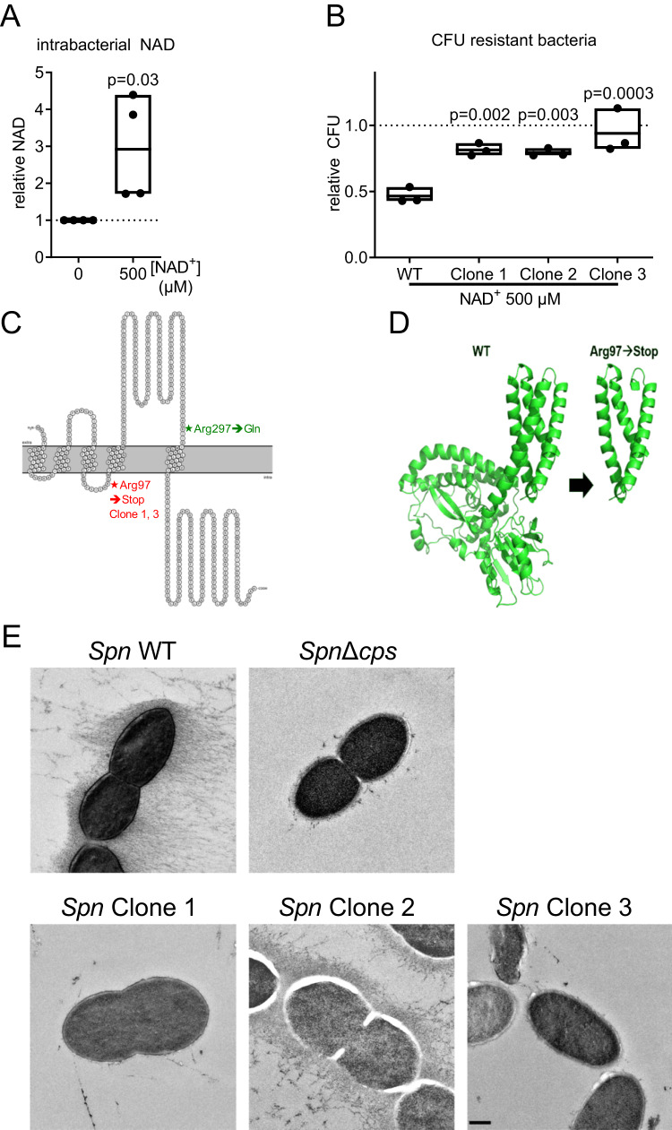
Fig. 6. Development of NAD+ resistance is associated with loss of capsule.
A Spn D39 was inoculated in a cell culture medium with and without NAD+ treatment as indicated. Bacteria were lysed 6 h post-inoculation and the total intracellular NAD was determined. B–D Spn D39 was cultivated in a cell culture medium and passaged 6 times with increasing concentrations of NAD+(50 µM to 5 mM) or without treatment. Afterwards, bacteria were plated on NAD+ (500 µM) supplemented agar plates. Individual clones were picked and characterized for NAD+ resistance and growth behaviour and sequenced. B Spn D39 WT and resistant bacteria were treated with 500 µM NAD+ for 6 h and bacterial counts were determined. C Spn D39 clones passaged with increasing concentrations of NAD+ and a control passaged in cell culture medium without NAD+ were sequenced. Identified amino acid changes in the protein CPS2E are displayed. Asterisks indicate amino acid substitutions. D 3D protein structure of CPS2E wild type and Arg97→Stop, generated with AlphaFold. E Transmission electron microscopy of Spn D39 WT, Δcps and the NAD+ resistant clones. Bacteria were cultivated until the early logarithmic phase in liquid media, cryo-fixated and the capsule stained using OsO4. Statistics: two-tailed paired t-test (A); One-way ANOVA with Fisher’s LSD (B); significance was determined against the untreated control (A) or the wild-type bacteria (B); Scale: 250 nm; N = 4 biologically distinct samples (A); 3 biologically distinct samples (B); box plots: line at mean; box ranges from min to max; results are normalized against untreated controls. WT wild type.

