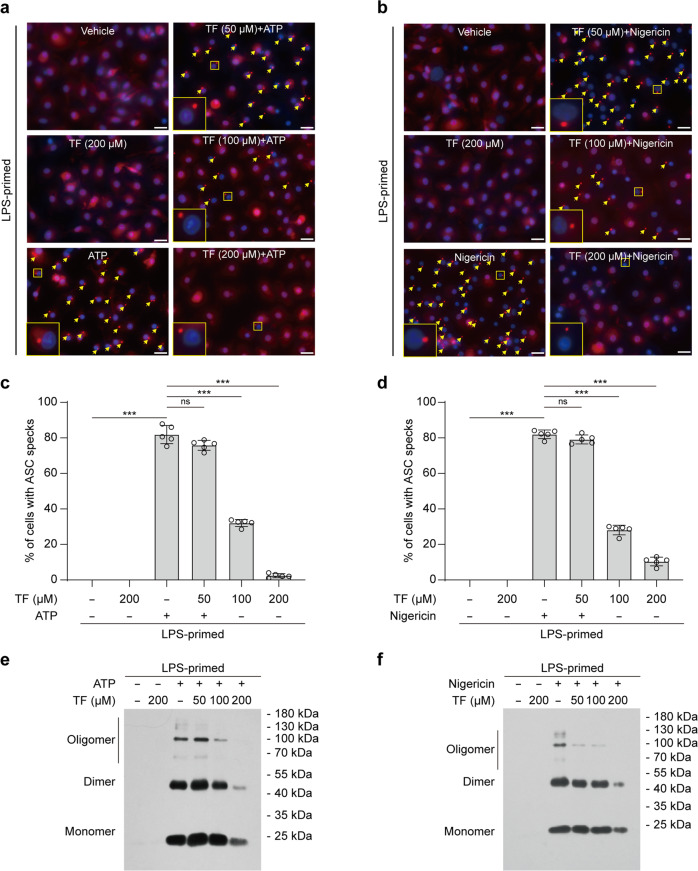
Fig. 2. Theaflavin blocks ASC speck formation and oligomerization upon NLRP3 activation.
BMDMs were primed with LPS (0.5 µg/mL) for 4 h and then pretreated with indicated doses of theaflavin for 1 h, followed by stimulation with ATP (4 mM) for 30 min (a, c, e) or nigericin (5 μM) for 1 h in the absence of LPS (b, d, f). Immunofluorescence microscopy analysis of ASC distribution (red) in BMDMs treated with ATP (a) or nigericin (b). Nuclei (blue) were revealed by Hoechst 33342 staining. A set of representative images are shown with ASC specks being indicated by yellow arrows. Scale bars, 20 μm. Percentages of cells with ASC specks were quantified in BMDMs treated with ATP (c) or nigericin (d). Western blot analysis of ASC oligomers in Triton X-100 insoluble, disuccinimidyl suberate-cross-linked pellets from BMDMs stimulated with ATP (e) or nigericin (f). Data are shown as mean ± SD (n = 5). ***P < 0.001; ns not significant, TF theaflavin.

