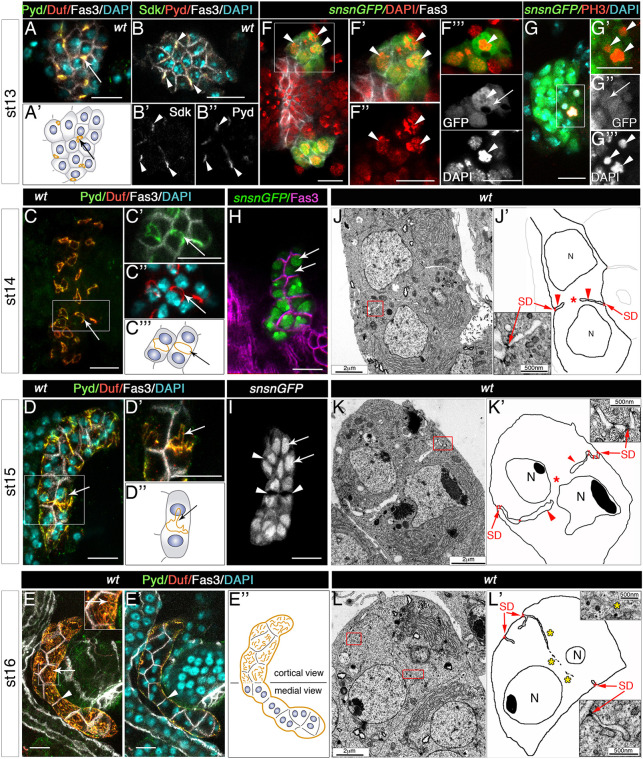
Fig. 1.
Genesis of nephrocyte slit diaphragms during embryogenesis. (A-E″) Confocal images and schematics of wild-type garland nephrocytes at developmental stages 13 to 16, stained with the indicated antibodies. Z-projections of stacks are shown except in B-B″, E′ (single planes). (B-B″) Sdk and Pyd colocalise in tricellular contacts [arrowheads, n=32 nephrocytes (N)/4 strings (S)]. Arrows in A,A′,C-E point to the accumulation of Duf and Pyd in tricellular contacts (A,A′, n=87N/5S) in a ring located at the equatorial cortex (C-C‴, n=124N/9S), which becomes distorted in D-D″ (n=137N/12S) and in a ruffled pattern on the nephrocyte outer surface (E, n=153N/8S). Arrowheads in E,E′ point to Fasciclin 3 (Fas3) accumulation in contact membranes between adjacent nephrocytes. (F-I) Single sections or z-projection (G) of sns-GCN-nGFP nephrocytes showing the presence of mitotic figures at stage 13 (arrowheads, F-F‴, n=32N/7S, DAPI staining; G-G‴, n=9N/6S, anti-PH3 staining) in cells that also show cytoplasmic distribution of nGFP (F‴ and G″, arrows); note the nuclear confinement of GFP in binucleated nephrocytes (arrows) at stages 14 (H, n=99N/7S) and 15 (I, n=71N/4S). White squares denote the regions magnified in adjacent panels; F-F″ and F‴ display the same cluster of nephrocytes at two different focal planes. Arrowheads in I show lumina of membrane invaginations. (J-L′) TEM micrographs and schematics of medial sections of stage 14 (J, n=12N), 15 (K, n=15N) and 16 (L, n=10N) wild-type garland nephrocytes. Red squares indicate enlarged regions in nearby panels, arrows point to slit diaphragms (SD), arrowheads to channels formed by membrane ingressions between sibling nuclei (N) and asterisks to cytoplasmic continuity (J′ and K′) or to membrane fragments (L′) located between the nuclei. Scale bars: 10 µm (A-I); 2 µm (J,K,L); 500 nm (J′,K′,L′).

