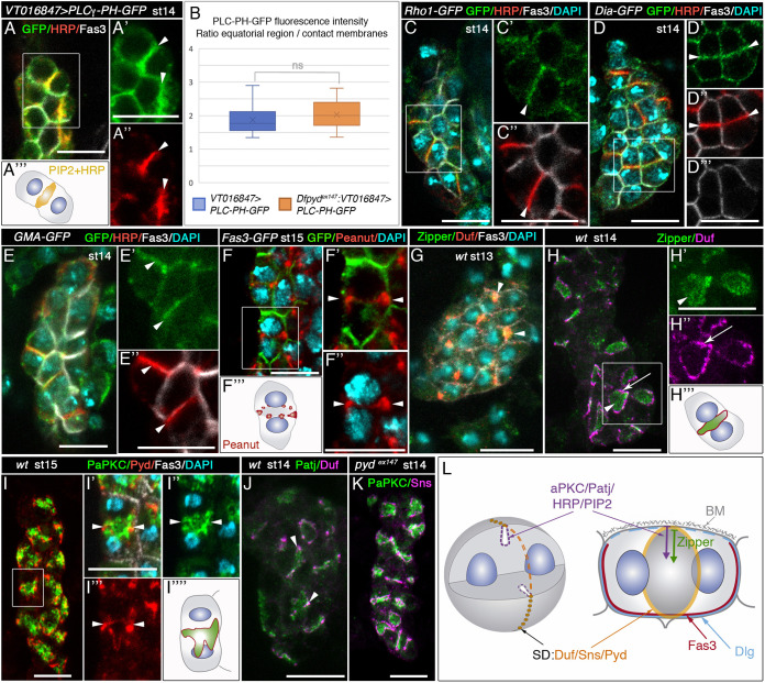
Fig. 3.
Acytokinetic cell divisions break membrane symmetry in embryonic garland nephrocytes by generating PIP2-enriched microdomains. (A-K) Single sections (A,C-E) and confocal z-projections (F-K) of embryonic garland nephrocytes of the specified developmental stages and genotypes, stained with the indicated antibodies. (A-A‴) In nephrocytes expressing PLCγ-PH-GFP, PIP2 accumulates at the equatorial plane, colocalising with the HRP epitope [arrowheads in magnified views A′,A″, n=12 nephrocytes (N)/2 strings (S), schematic in A‴]. (B) Quantification of PLCγ-PH-GFP fluorescence intensity per area in wild-type (Fig. 3A′) and pydex147 (Fig. 4C′) backgrounds (N=24/S=4 and N=21/S=6, respectively). Data show the ratio between the signal at the equatorial region and that at the site of contact between adjacent nephrocytes. Box plot shows median values (horizontal bars), the mean values (indicated by X) and the first to third interquartile ranges (boxes); whiskers extend to the minimum and maximum data values. ns, not significant. (C-E″) GFP-tagged versions of Rho1 (C,C′, n=25N/6S), Dia (D,D′, n=44N/3S) and GMA (E,E′, n=24N/7S) accumulate at the equatorial region of stage 14 nephrocytes, colocalising with the HRP epitope (arrowheads in C′,C″,D′,D″,E′,E″). Note, this region is devoid of Fas3 (C″,D‴,E″). (F-F‴) Peanut localises at the nephrocyte equatorial cortex (arrowheads in magnified views F′,F″, schematic in F‴, n=32N/4S). (G-H‴) Zipper mobilises from tricellular contacts, where it colocalises with Duf, in mononucleated nephrocytes (G, arrowheads, n=30N/2S) to the equatorial plane of binucleated nephrocytes (H,H′, arrowheads, n=77N/5S; see Fig. S4). Simultaneously, Duf accumulates at the equatorial cortex (H,H″, arrows, schematic in H‴; see also Fig. 2F-G′, Fig. S4). (I-J) PaPKC (I-I″, n=160N/11S) and Patj (J, n=45N/4S) accumulate at the equatorial region of binucleated nephrocytes, partially colocalising with Pyd (I) and Duf (J) at the cortex (arrowheads, I′-I‴,J). (K) PaPKC and Sns distribution do not change in the absence of slit diaphragms (pydex147 mutants, n=32N/3S). (L) Diagram showing the subcellular distribution in binucleated stage 14 nephrocytes of the indicated proteins and PIP2. Slit diaphragm components (in orange) accumulate in a ring at the equatorial cortex, Zipper (green arrow) only in the septum and apical determinants, the HRP epitope and PIP2 at the septum and at the equatorial cortex (purple arrow), colocalising in the latter region with slit diaphragm components. Scale bars: 10 µm.

