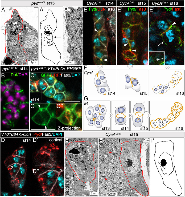
Fig. 4.
Establishment of PIP2-enriched membrane domains accelerates slit diaphragm assembly. (A-A″) TEM image, schematic and detail of stage 15 pydex147 nephrocytes [n=12 nephrocytes (N)/3 embryos]. Note the absence of equatorial membrane invaginations (black arrows) and slit diaphragms (compare with Figs 1K,K′ and 2D-G). (B) Confocal z-projection of pydex147 nephrocytes, showing Duf accumulation at the equatorial cortex [n=81N/5 strings (S)]. (C-C″) In pydex147 nephrocytes expressing UAS-PLCγ-PH-GFP (n=21N/6S), devoid of membrane invaginations, PIP2 accumulates in a ring in the equatorial cortex (C″, arrowheads in C′, quantification in Fig. 3B; C,C′ single sections). (D-D″) Overexpression of Ocrl induces the premature localisation of Pyd at the nephrocyte outer membrane (D′, cell 1, cortical view; D″, cell 2, medial view, n=29N/4S; compare with Fig. 1C-C‴,E-E″). (E-I′) Individual optical sections (E,E″), z-projection (E′), schematics (F,I′) and TEM images (H-I) of CycAC551 nephrocytes at the indicated embryonic stages, and schematic of control nephrocytes (G). When the extra acytokinetic cell division is prevented, Duf and Pyd accumulate at tricellular contacts between mononucleated nephrocytes at stages 14 (n=18N/3S) and 15 (n=18N/4S) (arrowheads in E,E′, compare with Figs 1A,C,D and 4G). At stage 16, Pyd still localises to cell contacts in some nephrocytes (E″, arrowhead; n=6N/3S), and scattered Pyd distribution can be observed at nephrocyte membranes exposed to haemolymph (arrows in E″, the inset shows a superficial view; compare with Fig. 1E). Nuclei (DAPI staining) are shown in blue in E-E″. Unlike the wild-type, stage 15 CycA nephrocytes lack slit diaphragms (H-I′), although occasionally electron-dense material is observed between adjacent nephrocytes (H′, red arrowheads). Scale bars: 2 µm (A,H,I); 500 nm (A″,H′); 10 µm (B-E″).

