
Fig. 3.
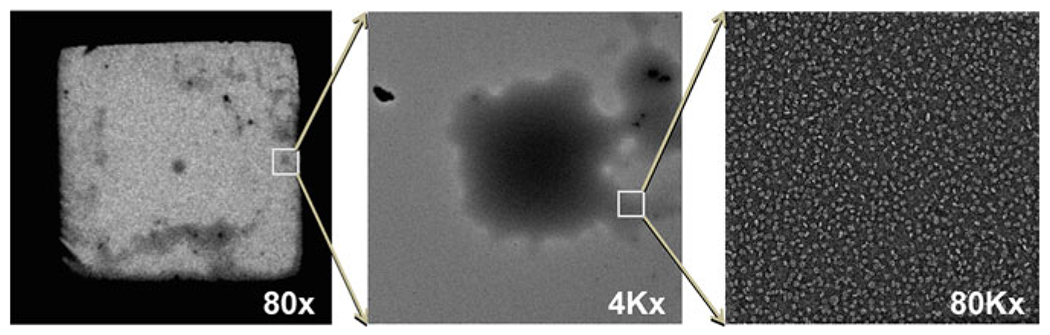
The best EM imaging area. The best imaging area of the protein was generally from the area that contained the thicker stain. Micrographs showing cloudy areas to locate and obtain images by EM for lipoproteins. Cloud (highlighted by box) of lipoprotein to designate lipoprotein location at 80× magnification (left), same designated area of cloud (highlighted by box) of lipoprotein further magnified at 4Kx (middle), and same designated cloud area further magnified at 80Kx (right).