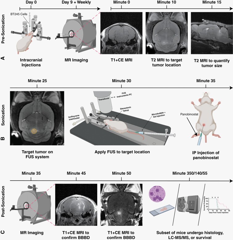
Figure 1.
Workflow for our MRI-guided FUS/MB treatments. (A) Prior to FUS treatment, patient-derived BT245 DIPG cells were injected i.c. into the pons region. Mice were placed in MRI and axial T1w images, and coronal and sagittal T2w images were acquired. (B) During FUS treatment, coronal T2w MRI images were used to coregister the FUS targeting system. Mice were then moved from MRI on the same bed to the FUS system, and a water tank was placed on the mouse head with ultrasound gel for acoustic coupling between the animal and ultrasound transducer. Microbubbles and gadolinium (Gd) contrast were injected i.v., the FUS transducer was stereotactically translated to align the acoustic focus to the pons region of the brain guided by the MRI image, and FUS was applied. Directly after FUS treatment, mice were injected with panobinostat i.p. (C) Post-FUS, mice were moved back to the MRI to assess BBB opening by T1w MRI scan of Gd extravasation. Mice were then sacrificed and assessed for histology and drug delivery (LC-MS/MS), or housing to continue survival studies. The timeline of study is shown above all images.

