Figure 6.
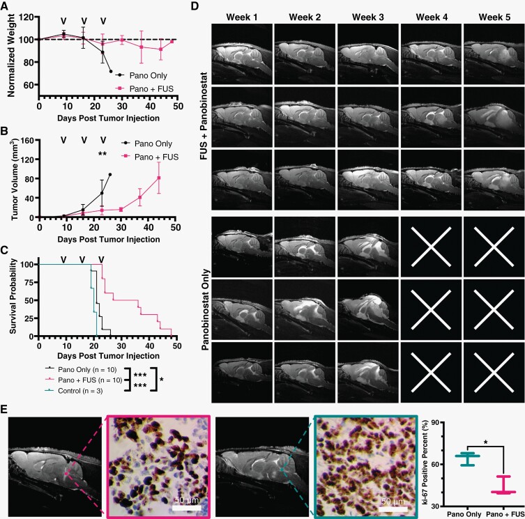
Focused ultrasound (FUS)/microbubbles (MB) with panobinostat reduces tumor growth and improves survival. (A) Longitudinal study of mouse body weight obtained weekly, (B) tumor volume using T2w MRI, and (C) KP survival curves. Black arrows on plots represent the treatment days. (D) Representative T2w MRI (sagittal axis) images of tumor progression for 3 representative mice in each group. Top 3 rows show MB + FUS with panobinostat-treated mice, and bottom rows are only panobinostat (no MB + FUS). White “x” illustrates the death of the mouse prior to the MR imaging that week. (E) Representative images of Ki-67 staining and its relation to MR images (left) and quantification of Ki-67 positive cells in tumor region for panobinostat + MB + FUS and panobinostat only (right). Significance testing was done using an unpaired Student’s t test for B (n = 10) and E (n = 3), and Mantel-Cox test for C (n = 10), where *, **, *** indicate P < .05, P < .01, and P < .001, respectively.

