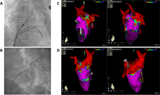
Fig. 2.
An overview of the index procedure (panel A and B), of a 3D mapping pre-ablation during the redo (left side panel C and D) and after radiofrequency ablation (right side panel C and D). Pulsed field ablation of the left inferior pulmonary vein in basket (panel A, AP projection) and flower (panel B, LAO 60°) configuration using the Farawave 31-mm catheter, a decapolar catheter in the coronary sinus and a quadripolar catheter in the right ventricle. Panels C and D: Multielectrode mapping of the left atrium at the repeat procedure revealing gaps in the left carina (panel C, left lateral view, before and after ablation), right anterior carina and posterior aspect of the right inferior pulmonary vein (panel D, right lateral view, before and after ablation). Voltage thresholds used: 0.2 and 0.5 mV

