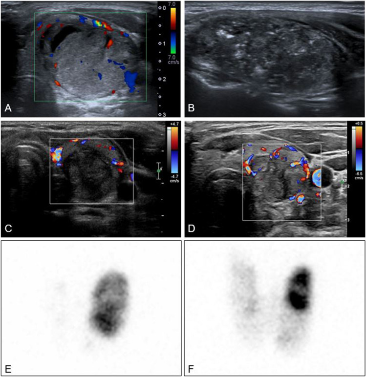
Fig. 2.
A–D Colour Doppler and grey scale ultrasound images of a patient successfully treated with RFA for STA. A Before RFA, showing a hypervascular nodule, volume 13.9 mL. B During RFA procedure with typical hyperechoic zones. C 4 months after RFA, volume 3.9 mL. The patient is biochemically euthyroid. No vascularity visible in treated zone but some vascularity in periphery of nodule. D 2 years after RFA treatment. Volume remains 4.0 mL and patient continues to be euthyroid. 2E-F 123I scintigraphy image of thyroid. E Before RFA depicting a left sided hot nodule with near complete suppression of thyroid tissue. F 15 months after RFA. Reduction of volume and autonomy of left sided nodule and increased uptake of thyroid tissue

