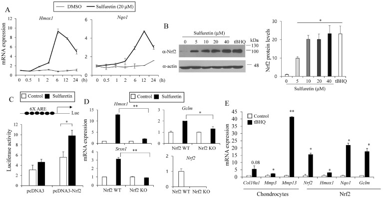
Fig. 2.
Sulfuretin stimulates Nrf2 activity. (A) C3H10T1/2 cells were treated with sulfuretin (20 μM) for 0.5, 1, 2, 6, 12, and 24 hours and relative temporal expression levels of Hmox1 and Nqo1 were measured by real-time PCR. (B) Sulfuretin treatment at 5-40 μM for 6 hours in C3H10T1/2 cells increased Nrf2 protein levels as determined by western blotting. Actin was used for loading control and tBHQ (50 μM) was used as a control. Representative blot images and relative densitometry bar graph were shown (n = 3). (C) HEK293T cells were transfected with the Nrf2 dependent-antioxidant response element (ARE) luciferase reporter construct (6XARE) and an expression vector coding for Nrf2 (pcDNA3-Nrf2). After 24 hours, cells were treated with DMSO (control) or sulfuretin (20 μM) for 24 hours and luciferase activity was measured. Luciferase signals were normalized with Renilla luciferase activity. (D) Nrf2 wild type (WT) MEF or Nrf2 knockout (KO) MEF were treated with sulfuretin (20 μM) for 12 hours and expression levels of Nrf2 target genes were measured by real-time PCR. (E) C3H10T1/2 cells were differentiated into chondrocytes in the presence of tBHQ (100 μM) and relative levels of hypertrophic genes and Nrf2 target genes were determined by real-time PCR. Data are presented as mean ± SEM (n = 3). Statistical significance was determined by comparison to the control using Student’s t-test (*P < 0.05; **P < 0.01).

