
Figure 4.
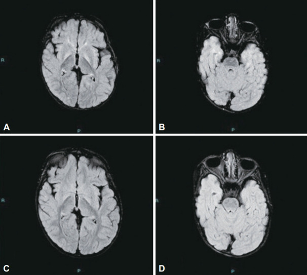
FLAIR axial images show bilateral, symmetrical mildly increased signal intensity changes along the globus pallidus (A) and medial aspect of both temporal lobes (B) with no diffusion restriction in kernicterus. FLAIR axial images of follow-up magnetic resonance imaging show partial regression of the bilateral symmetrical increased signal intensity along the medial globus pallidus (C) and the medial temporal lobes (D), representing radiological improvement in postkernicterus changes. FLAIR, fluid attenuated inversion recovery.