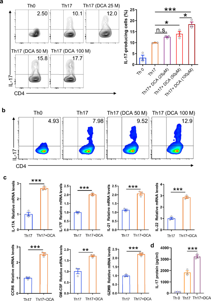
Fig. 1.
DCA promotes Th17 cell differentiation. a Splenocytes were differentiated under Th0 or Th17-polarizing conditions for 72 h in the absence or presence of different dosage of DCA, then the cells were re-stimulated with PMA/ionomycin and analyzed for the percentages of IL-17-producing CD4+ T cells by flow cytometry (n = 3). b Naïve CD4+ T cells were differentiated under Th0 or Th17-polarizing conditions in the absence or presence of DCA. Cells were then re-stimulated with PMA/ionomycin and the percentages of IL-17-producing CD4+ T cells were determined. c Naïve CD4+ T cells were differentiated under Th17-polarizing conditions for 72 h in the absence or presence of DCA (100 µM), then the cells were re-stimulated with PMA/ionomycin for 5 h and mRNA expression levels of indicated genes was detected by real-time PCR (n = 3). d Naïve CD4+ T cells were differentiated under Th0 or Th17-polarizing conditions for 72 h in the absence or presence of DCA (100 µM). Cells were then re-stimulated with PMA/ionomycin for 12 h and the supernatants were analyzed for IL-17 by ELISA (n = 3). *p < 0.05; **p < 0.01; ***p < 0.001. n.s.: no statistically significant difference (p > 0.05). One-way ANOVA with Tukey's multiple comparisons tests (a right panel, d) or two-tailed Student’s t-tests were used (c). Data are expressed as mean ± SEM from at least three independent experiments or representative data (a: left panel, b)

