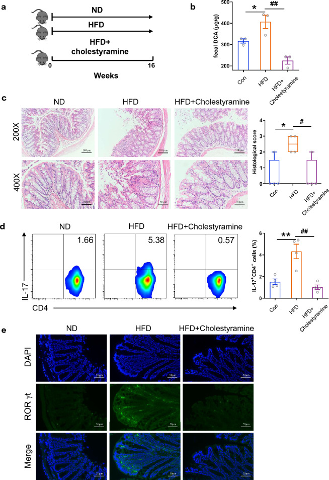
Fig. 6.
Cholestyramine treatment alleviates HFD-associated colonic inflammation. a Animal treatment procedure (ND, n = 5; HFD, n = 6; HFD + VCM, n = 7). b Fecal DCA concentrations (n = 3). c Representative HE staining and histological score of colon sections from ND, HFD and HFD plus cholestyramine treated mice (n = 4). Scale bar, 100 µm (×200) and 50 µm (×400). d The percentage of IL-17-producing cells from mesenteric lymph nodes of ND, HFD and HFD plus cholestyramine treated mice (n = 4). e Representative RORγt (green) and DAPI (blue) immunofluorescence staining of colon tissues from ND, HFD and HFD plus cholestyramine treated mice (Scale bar, 50 µm). *p < 0.05; **p < 0.01 compared to the normal diet control mice. #p < 0.05; ##p < 0.01 compared to the HFD plus cholestyramine treated mice. One-way ANOVA with Newman-Keuls multiple comparisons test (b) or Tukey's multiple comparisons tests (c, d) were used. Data are expressed as mean ± SEM from at least three independent experiments or representative data (c and d: left panel, e)

