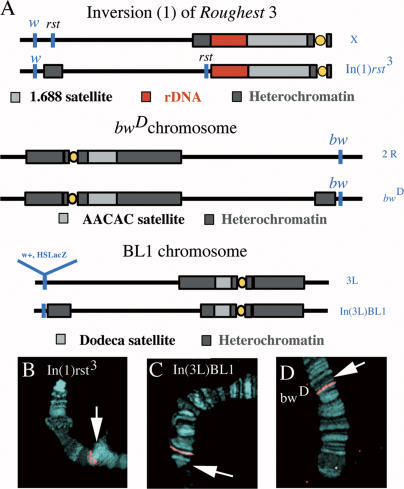
Figure 9. Variegating Lines and Probes Used in This Study.
Variegating lines were used for three different chromosomes, each variegating for a different gene. For full details see Materials and Methods.
(A) Diagram of each line showing approximate breakpoints and locations of variegating genes. FISH probes were made from P1 clones covering each gene and from heterochromatic sequences unique to each chromosome (see Materials and Methods).
(B–D) FISH probes for each chromosome. FISH probes were hybridized to polytene squashes to show cytological location of each probe. Regions of proximal heterochromatin from inversion and insertion are marked with an arrow. Each chromosome spread is taken from individual experiments. (B) Probe used in experiments for the line In(1)rst3. (C) Probe used in experiments for the line In(3L)BL1. (D) Probe used in experiments for the line bwD.

