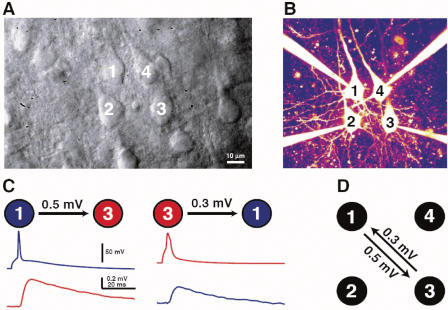
Figure 1. Illustration of a Quadruple Whole-Cell Recording.
(A) Dodt contrast image showing four thick-tufted L5 neurons before patching on.
(B) Fluorescent image of the same four cells in whole-cell configuration.
(C) Average EPSP waveform measured in the postsynaptic neuron (bottom) while evoking action potentials in the presynaptic neuron (top).
(D) Diagram of detected synaptic connections and their strengths for this quadruple recording.

