Figure 1.
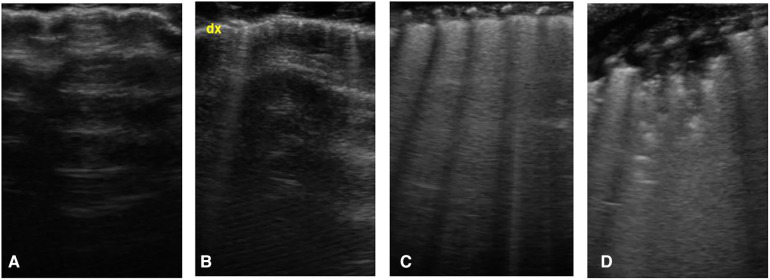
Lung ultrasound score, images obtained by longitudinal scan with a linear probe: (A) presence of only A-lines under the pleural line (normal pattern, score 0); (B) isolated vertical bright line, starting from pleural line and erasing the underlying A-lines, called B-line (score 1); (C) coalescent B-lines (score 2); (D) presence of consolidation (score 3).

