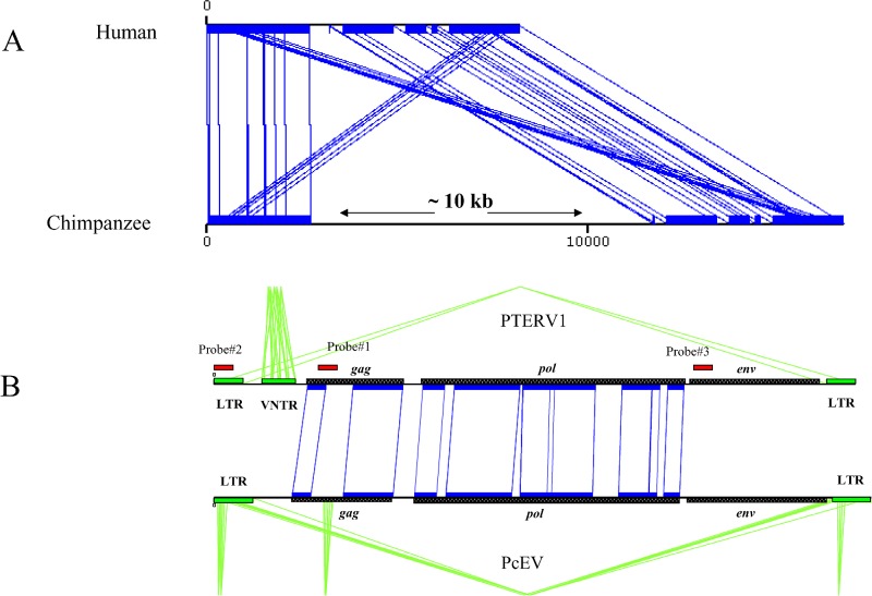
Figure 1. Identification and Sequence Analysis of PTERV1.
(A) A graphical alignment of chimpanzee genomic sequence (AC097267) and an orthologous segment from human Chromosome 16 (Build 34) depicting an example of a PTERV1 (approximately 10 kb) insertion. Aligned sequences are shown in blue (miropeats) [47].
(B) The typical retroviral structure of the insert (gag, pol, env, and LTR) is compared to a baboon (Papio cynocephalus) endogenous retrovirus (PcEV). Regions of nucleotide homology are designated by black blocks and inter-sequence connecting lines. The location of probes (see Table S1) used in genomic library hybridizations, Southern blot analyses, and neighbor-joining tree analyses are shown (red).

