Figure 3.

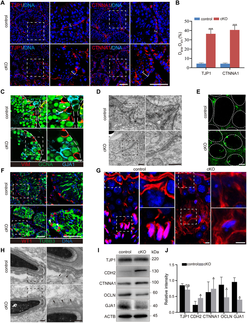
Deletion of Pik3c3 in Sertoli cells resulted in impaired Sertoli cell polarity. (A) Immunofluorescence of TJP1 or CTNNA1 in control and cKO mice testis at 8 W of age. Bar: 50 μm. (B) Graph showing the distance traveled by TJP1 or CTNNA1 beyond the BTB near the basement membrane (DTJP1 or DCTNNA1) versus the radius of the seminiferous tubule (DSTr) per seminiferous tubule. The largest cross-section of each testis was used for staining and cell counting (n = 6). ***P < 0.01. (C) Immunofluorescence of VIM, GCNA and GJA1. Filled triangles, gap junctions localized between Sertoli cells and germ cells. Bars: 10 μm. (D) TEM showed the BTB structure in control and cKO testis. Filled triangles, electron rich areas referred as tight junctions. Bars: 400 nm. (E) Immunofluorescence of FITC showed the disturbed BTB structure in cKO testis. FITC was injected into the control and cKO mice and testes were isolated 1 h later for cryosection. Bar: 50 μm. (F) Immunofluorescence of WT1 and TUBB3. Filled triangles, Sertoli cell nuclei traveled away from the basement membrane of seminiferous tubule. Bar: 50 μm. (G) Immunofluorescence of phalloidin. Bar: 5 μm. (H) TEM of aES structure. Filled triangles, actin bundles. Bar: 300 nm. (I-J) Immunoblotting of TJP1, CDH2, CTNNA1, OCLN and GJA1 proteins. The expression of ACTB was used as internal control (I). The relative intensity of TJP1, CDH2, CTNNA1, OCLN and GJA1 was shown as compared to the expression of ACTB (J).
