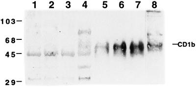
FIG. 3.
Effect of RFP treatment on CD1b in membrane and cytosol fractions. Cells were treated with GM-CSF plus IL-4 (lanes 1 to 3 and 5 to 7) in the presence of RFP at 2 (lanes 2 and 6) or 10 (lanes 3 and 7) μg/ml. Cell homogenates were separated into membrane and cytosol fractions. Each fraction (20 μg) was separated by SDS-polyacrylamide gel electrophoresis and visualized by immunoblotting with the polyclonal antibody against CD1b. Lanes 1 to 4, cytosol fractions from AMNC (lanes 1 to 3) and C1R/CD1b cells (lane 4); 5 to 8, membrane fractions from AMNC (lanes 5 to 7) and C1R/CD1b cells (lane 8). The values on the left are molecular size standards (in kilodaltons).

