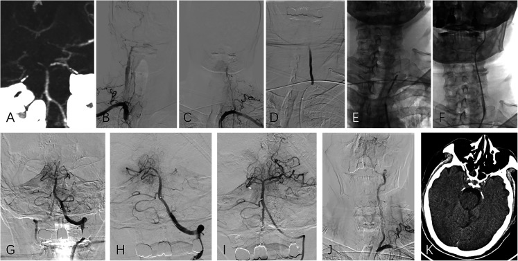
Figure 3.
(Case No. 8), dominant left vertebral artery occlusion with non-dominant right vertebral artery occlusion (type C tandem occlusion). (A) CT angiography showed basilar artery tip occlusion and dominant vertebral artery at left side. (B, C) Bilateral subclavian arteries angiography depicted left vertebral artery occlusion and a slender right vertebral artery. (D) A 300 cm 0.014-inch microguidewire, with a 0.021-inch microcatheter form a coaxial system and was used to pass through the stenosis/occlusion segment. (E, F) After confirmation of the true cavity with microcatheter angiography, a balloon (3 mm diameter) was introduced to dilate the stenotic segment, and the guiding catheter was then advanced to the V2 segment through the stenotic segment over the partially inflated balloon. (G-I) Basilar artery thrombectomy was performed with a combination of stent retriever and aspiration. (J) After confirming intracranial successful revascularization, the microguidewire was sent to the V2 segment, and the guiding catheter was pulled back with gentle aspiration to remove all possible residual intraarterial thrombi. A balloon-expandable stent was then implanted at the ostial vertebral artery over the microguidewire. (K) The follow-up CT showed a small infarction in the brain stem.

