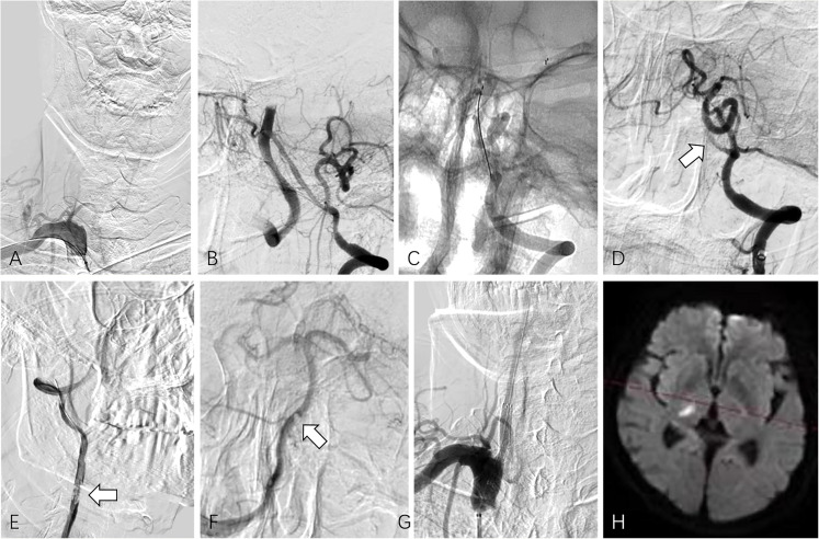
Figure 4.
(Case No. 7), dominant right vertebral artery occlusion with non-dominant left vertebral artery patency (type A tandem occlusion). (A) Selective angiogram of the right subclavian artery depicted right vertebral artery occlusion. Initially, attempts to pass the microguidewire through the occlusion segment of the right vertebral artery failed. (B) Left vertebral artery angiography showed the small size of the V4 segment of the left vertebral artery. (C) Mechanical thrombectomy for basilar artery with Solitaire AB stent retriever was made via the left vertebral artery for two times. (D) After each thrombectomy, angiography showed the occlusion of the left V4 segment (arrow). (E) A coaxial system consisting of a 5 Fr MPA catheter and a 0.035-inch hydrophilic coated guidewire was used to successfully pass through the occluded segment of the right vertebral artery, and an angiogram showed the residual intraarterial thrombi (arrow). (F) Right vertebral artery angiogram showed patency of basilar tip, and a filling defect was detected at the vertebrobasilar junction (arrow), which should be the thrombus stuck at the left vertebral artery. (G) After removal of residual thrombus by aspiration during catheter withdrawal, a self-expendable stent was implanted at the stenotic segment of the right vertebral artery over the microguidewire. (H) Follow-up MRI revealed a small infarction in the right thalamic region.

