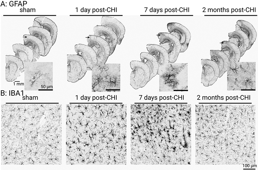
Figure 2: The temporal patterns of astrocyte (GFAP) and microglia (IBA1) morphological changes after a CHI.

(A) GFAP staining at low magnification shows the regional increase in staining seen in the cortex of the CHI group. The morphological appearance of the astrocytes is shown in the higher-magnification insets, which were taken from the middle brain sections and from the same regions of the cortex. (B) IBA1-positive staining in the cortex at 1 day, 7 days, and 2 months post-injury shows changes in microglia morphology in the neocortex after the CHI (n = 7-14, 50/50 male/female). The mice (CD-1/129 background) were 8 months old at the time of surgery. This figure has been adapted from 11 and reproduced with permission. Scale bar = 1 mm, 50 μm and 100 μm as indicated in the figure.
