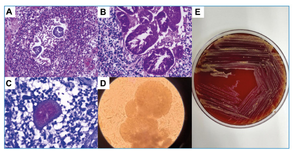
FIGURE 1: Microbiological and histopathological findings; (A) Grain in hematoxylin and eosin stain; (B) Grain in periodic Acid-Schiff stain; (C) Grain in Fite-Faraco stain; (D) Direct microcopy with 40% potassium hydroxide showing white homogenous grain; (E) Colonies of Gordonia soli in blood agar medium culture after 72 h of incubation at 30ºC.

