Fig. 3.
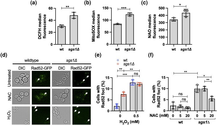
Endogenous ROS, mitochondrial mass, and recombinogenic DNA lesion formation in the sgs1Δ mutant. a) Measurement of ROS in the sgs1Δ mutant by flow cytometry using the fluorescent dye DCFH diacetate. b) Measurement of mitochondrial superoxide content in the sgs1Δ mutant by flow cytometry using MitoSOX Red fluorescent dye. c) Measurement of mitochondrial mass in the sgs1Δ mutant by flow cytometry using the fluorescent dye NAO. d) To detect recombinogenic DNA lesions, homologous recombination factor Rad52 was tagged with GFP (Rad52-GFP), and foci formation detected by fluorescence microscopy (BZ-X170, Keyence) of exponentially growing cell cultures either untreated or treated with the antioxidant NAC (20 mM) for 2 hours or H2O2 (0.5 mM) for 30 minutes. Representative images are shown with arrows pointing to nuclei with Rad52-GFP foci. In nuclei without Rad52-GFP foci, the GFP signal appears diffused throughout the nucleus. e) Percentage of cells with Rad52-GFP foci in cultures of wildtype cells and the sgs1Δ mutant in the presence or absence of oxidative stress induced by H2O2. Exponentially growing cell cultures were incubated at 0.5 mM H2O2 for 30 minutes and imaged by fluorescence microscopy (BZ-X170, Keyence). f) Effect of the antioxidant NAC (5 mM, 20 mM) on the percentage of cells with Rad52-GFP foci in cultures of wildtype cells and the sgs1Δ mutant. Exponentially growing cell cultures were incubated at the indicated concentration of NAC for 2 hours and imaged by fluorescence microscopy (BZ-X170, Keyence). Experiments were performed in triplicate and the mean ± SD is reported. Statistical significance of differences was determined with a Student's t-test and reported as *P < 0.05; **P ≤ 0.01; ***P ≤ 0.001; ns, not significant.

