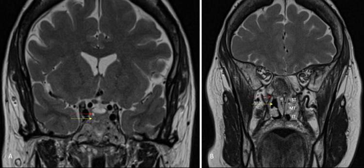
FIG. 2.
Postoperative coronal T2-weighted magnetic resonance image (A) showing the 2 × 2 mm hyperintense nodule (red arrow) and the resection tract, seen as a T2 hyperintensity, inferomedial to the nodule (yellow arrow). Coronal T2-weighted magnetic resonance image (B) at the level of the inferior orbital fissure showing the SPF (red arrow) adjacent to the site of the right middle and partial superior turbinectomy (yellow arrow). IT = inferior turbinate; MT = middle turbinate; ST = superior turbinate.

