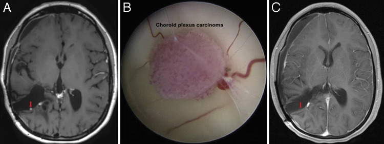
FIG. 2.
A: Preoperative axial postcontrast T1–sampling perfection with application optimized contrasts using different flip angle evolution (SPACE) MRI sequence showing focal punctate enhancement along the posterior wall of the patient’s prior right temporoparietal resection tract confirmed as local CPC recurrence. The third ventricle is notably free of disease. B: Endoscopic view of patient’s CPC recurrence in the previous right temporoparietal resection tract. The 2.5-mm tumor is multilobulated and hypervascular with surrounding vessels and attachment to parenchymal tissue at its base. C: Postoperative axial postcontrast T1-weighted MRI demonstrating absence of enhancement at the resection site, indicative of GTR. Vascular enhancement is noted adjacent to resected tumor site.

