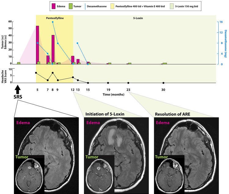
FIG. 1.
Case 1. Timeline of symptoms, imaging findings, and steroid dose according to treatment modality (upper). Axial T1-weighted sequences with contrast and FLAIR sequences at key time points (lower). Baseline MRI at the time of SRS (left) to a 1.8-cm falcine meningioma shows no edema. At 12 months (center), 5-Loxin was initiated after trimodal therapy failed to control AREs (imaging changes include central necrosis, rim of parenchymal enhancement, bifrontal edema). At 23 months (right), MRI shows resolution of the AREs (no perilesional edema, tumor volume decreased 61%). bid = twice a day; tid = three times a day; qid = four times a day; VAS = visual analog scale.

