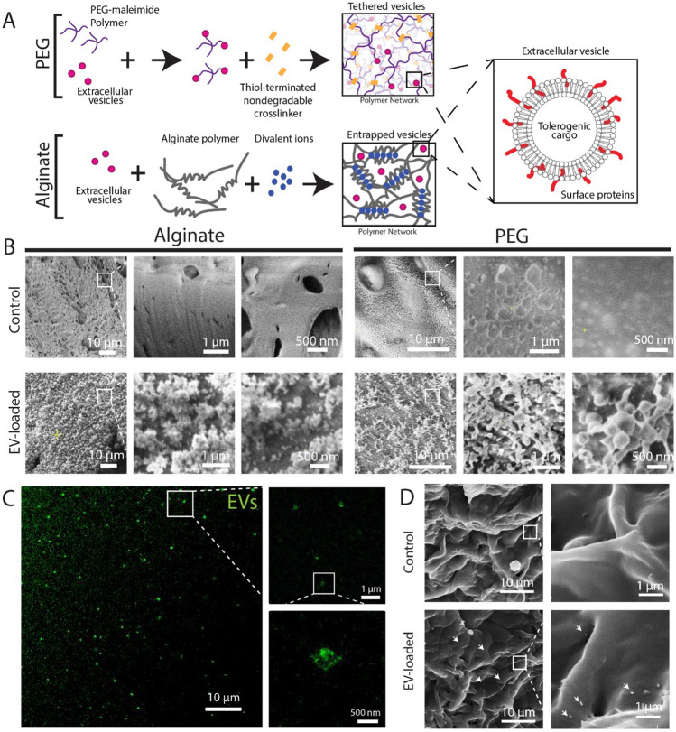
Fig. 4.
Imaging of EV-loaded hydrogel delivery systems. A Schematic illustrating EV tethering to PEG hydrogels and EV entrapment within alginate. B Cryo-scanning electron microscopy of blank (control, top row) and EV-loaded (bottom row) alginate (left) and PEG (right) hydrogels at increasing magnifications. C Stimulated emission depletion confocal microscopy of EV-loaded PEG hydrogels. D SEM imaging of PEG hydrogel surface. White arrows indicate EV location

