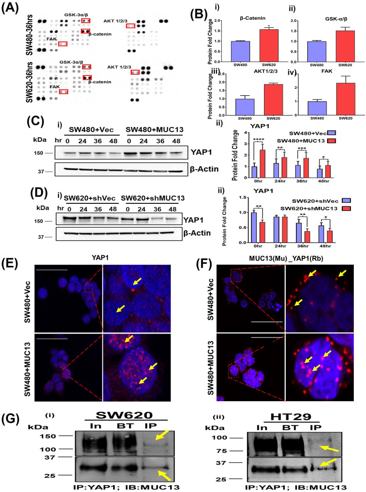
Figure 4. MUC13 and YAP1 proteins colocalize and interact during anoikis-induced conditions.
(A) Human kinase array analysis with the high (SW620) and low (SW480) MUC13-expressing isogenic cell lines after 36 h of anchorage-independent stimulation. The box indicates the differential expression of kinases with increased (red) and decreased (blue) expression. (B) Quantitation of the protein fold change of total β-catenin, GSK3-α/β (S21/S9), AKT1/2/3(T308), and FAK(Y397) phosphorylation. (C, D) Immunoblot analysis of YAP1 protein using MUC13 overexpressing knockdown cells. (C, D) Quantification of YAP1 expression in (C) SW480+Vector and SW480+MUC13, and (D) SW620+shVector and SW620+shMUC13. Data are representative of at least three individual experiments. The data are shown as mean ± SEM. Sidak’s multiple comparison test is used after a two-way ANOVA. *P < 0.05, **P < 0.01, ***P < 0.001, and ****P < 0.0001 denote significant differences. MUC13 overexpressing and vector control stable cell lines 36 h post anoikis induction, pelleted, cryofixed, and cryosectioned on slides. (E) Confocal imaging using YAP1 antibody in MUC13 overexpressing show higher YAP1 localization in the nucleus compared with vector-only control (scale bar, 50 μm). (F) PLA was performed using indicated anti-mouse (MAb) and anti-rabbit (PAb) antibodies along with PLA probes. Red dots indicated by yellow arrows represent the physical interaction among listed proteins in used cell lines. MUC13 expression influenced the interaction of YAP1 along with their nuclear translocation. The images depict at least three independent experiments (scale bar, 50 μm). (G) (i, ii) YAP1 and MUC13 physically interact in CRC cells. Immunoprecipitation of YAP1 in SW620 (i) and HT29 (ii) cells were able to pull down MUC13, as evidenced by immunoblot for MUC13 with I.P. of YAP1. Two different fractionated forms of MUC13 are indicated.

