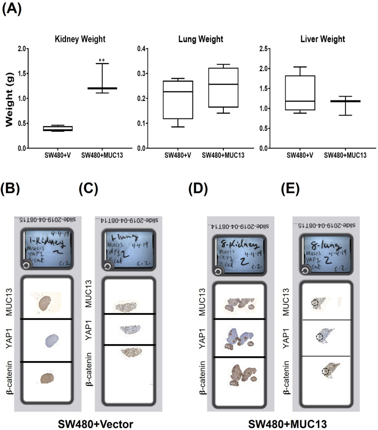
Figure S6. Tail vein metastasis model in NSD mice.
(A) Wet weight of mice’s three most affected organs, namely the kidney, lungs, and liver, injected with SW480+MUC13 and SW480+Vector cells. Organ weight profile of the kidney, lungs, and liver of mice injected with control and MUC13 overexpressing cells. (B, C, D, E) Digitally scanned complete slides of kidney and lung tissues collected from a metastatic mouse model and injected with SW480+Vec (B, C) and SW480+MUC13 (D, E) cells via tail vein. The harvested tissues were stained for MUC13, YAP1, and β-catenin using their respective antibodies. The data are represented as mean ± SEM. Sidak’s multiple comparison test is used after a two-way ANOVA. *P < 0.05, **P < 0.01, ***P < 0.001, and ****P < 0.0001 denote significant differences.

