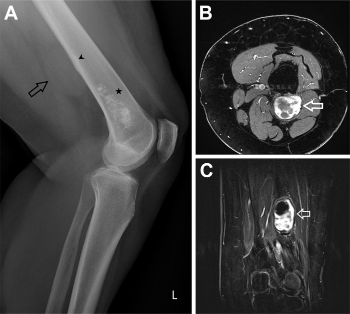
Fig. 1.
(A) Radiograph Knee lateral view. Black star: chondroid lesion in the distal femur. Black arrow: Soft tissue lesion visible without any calcifications in the lesion. Black arrowhead: Smooth regular thickening of the endosteum. Discreet periosteal reaction, single layered. (B) MRI T1 TSE fat saturated after Gadolinium transversal view. White arrow: After Gadolinium heterogenous uptake of the contrast agent, the T2 hyperintense areas do not enhance, only the T2 hypointense areas show enhancement. (C) MRI T1 TSE subtraction after Gadolinium coronal view. White arrow: After Gadolinium the hypointense border shows discreet peripheral enhancement. Heterogenous enhancement of the lesion, the T2 hyperintense areas do not enhance.

