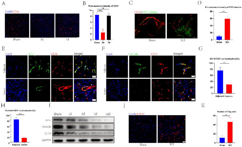
Fig. 1.
BSCBS and TJs were destroyed, and Treg cell infiltration increased after spinal cord injury. A, B The distribution and quantification of blood vessels labeled with CD31 (n = 3). C, D Fluorescence intensity and quantification of FITC-dextran around CD31-labeled blood vessels on day 7 after spinal cord injury (n = 4). E–H co-immunostaining and quantification of TJS-related proteins and vascular at injury sites and adjacent sites on day 7 after spinal cord injury (arrows: intact blood vessels; triangles: damaged blood vessels) (n = 3). I Western blotting detected the expression of TJs protein at each time point after spinal cord injury. J, K Representative staining and quantification of Foxp3-labeled Treg cells at the injured site after spinal cord injury (n = 3)

