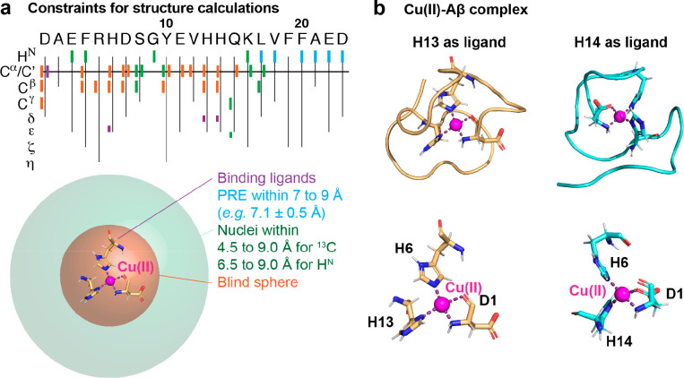
Figure 2.
Molecular structures of the Cu(II)-Aβ complex using paramagnetic NMR experiments. (a) Structural constraints from different paramagnetic NMR experiments where binding ligands (violet) are within the blind sphere of paramagnetic NMR experiments (orange). Nuclei detected with paramagnetic NMR but not with diamagnetic NMR pulse sequences are located within the decreased blind sphere of paramagnetic compared to diamagnetic experiments (green). PRE measurements provide additional structural constraints (cyan). (b) Molecular structures of two different binding modes with H13 or H14 as the fourth binding ligand (available as PDB structure 8B9Q or 8B9R, respectively). The assigned binding ligands are the nitrogen of the NH2 terminus, the amide oxygen of D1, the Nε of H6, and the Nδ of H13 or H14. Data were reproduced and the figure was adapted with permission from ref (1). Copyright 2022 the authors. American Chemical Society.

