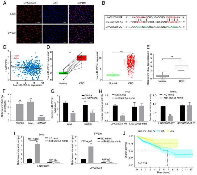
Figure 3.
LINC02038 sponges miR-552-5p in the CRC cells. (A) LINC08038 localized to the cytoplasm (red) and nucleus (blue) in LoVo and SW620 cells using FISH test. (B) The predicted binding site between LINC02038 and miR-552-5p. (C) Using data obtained from TCGA-COAD dataset, it was shown there was a negative association between LINC02038 and miR-552-5p expression in CRC tissues. (D) TCGA-COAD database analysis confirmed that miR-552-5p expression was higher in CRC tissues than in matched and unpaired normal tissues. (E) RT-qPCR analysis of miR-552-5p expression in CRC and normal tissues. (F) RT-qPCR detection of miR-552-5p expression in CRC cells. (G) miR-552-5p expression in LoVo and SW620 cells transfected with LINC02038 overexpression or an empty vector was determined using RT-qPCR. (H) The relative luciferase activity in LoVo and SW620 cells co-transfected with LINC02038 WT or MUT and miR-552-5p mimics. (I) RIP assays were used to examine LINC02038 enrichment in LoVo and SW620 cells co-transfected with LINC0203 and miR-552-5p mimic. (J) The relationship between miR-552-5p and overall survival in CRC patients in TCGA-COAD dataset. **P<0.01, ***P<0.001. miR, microRNA; CRC, colorectal cancer; FISH, Fluorescence in situ hybridization; TCGA, The Cancer Genome Atlas; COAD, colon adenocarcinoma; RT-qPCR, reverse transcription quantitative PCR; MUT, mutant; WT, wild-type; Ago2, argonaute 2; RIP, RNA immunoprecipitation.

