
Fig. 4.
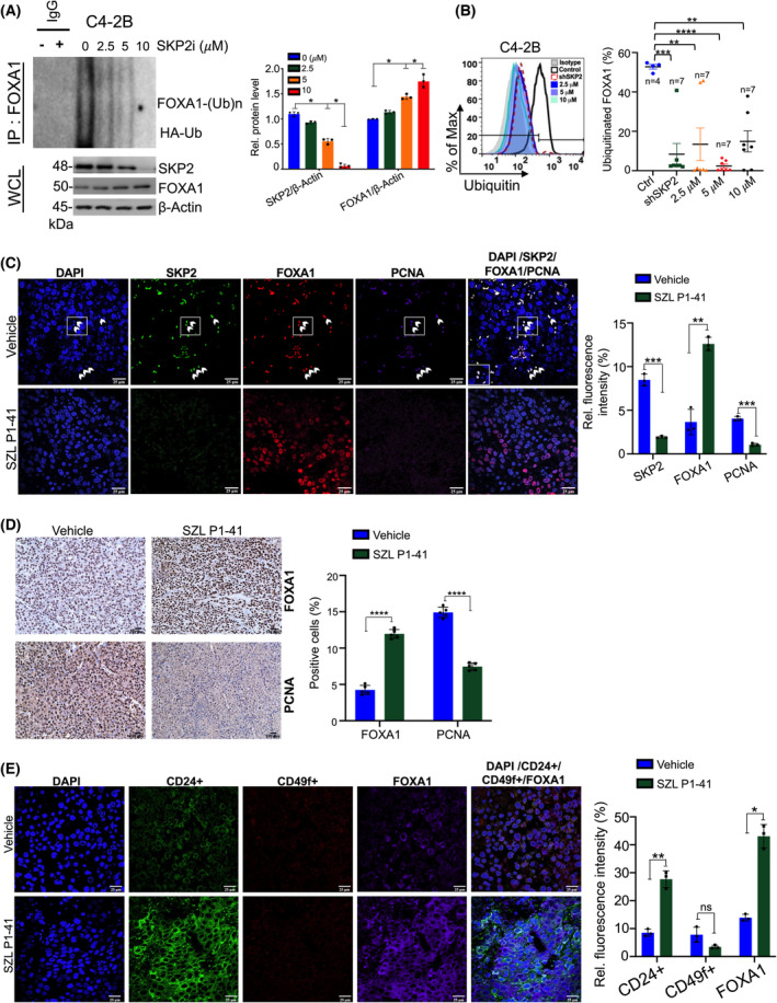
SKP2 alters FOXA1 ubiquitination levels in human prostate cancer cells in a dose‐dependent manner. (A) Endogenous ubiquitination of FOXA1 in C4‐2B proceeding SKP2 inhibition using SZL P1‐41 at different concentrations (μm). Samples were collected and subjected to in vivo ubiquitination and western blot analysis (n = 3). (B) FOXA1 ubiquitination profile using flow cytometry for vehicle, SKP2 KD, and SKP2 inhibition using SZL P1‐41 for C4‐2B. Representative histogram and percent ubiquitination of FOXA1 are plotted. Colored points represent number of replicates per group. (C) C4‐2B xenograft mice received vehicle (DMSO) or SKP2 inhibitor SZL P1‐41 (30 mg·kg−1; three times per week, intraperitoneal, i.p. injection) for 30 days. Immunofluorescence (IF) staining displays increased colocalization (white arrows) amongst SKP2, FOXA1, and PCNA for C4‐2B xenograft vehicle tissues, while exposure to SZL P1‐41 treatment reduced SKP2 and PCNA levels (n = 3). Scale bars are 25 μm. (D) C4‐2B xenograft tissue sections were subjected to Immunohistochemistry (IHC) staining with the indicated antibodies (n = 5). Scale bars are 100 μm. (E) IF luminal (CD24+) and basal (CD49f+) lineage staining in C4‐2B vehicle and SZL P1‐41‐treated mice (n = 3). Scale bars are 25 μm. Comparison between groups was performed using Student's t‐test. Bars indicate SEM. *P < 0.05, **P < 0.01, ***P < 0.001, ****P < 0.0001.