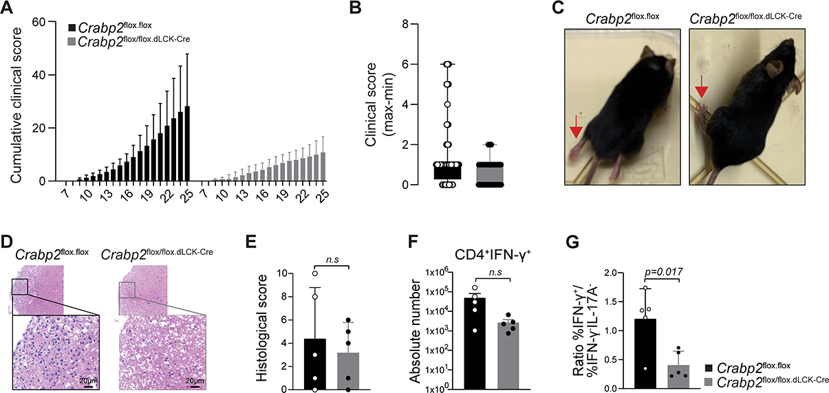
FIGURE 7: Extranuclear RARα signaling controls effector differentiation in vivo. (Related data shown in Figure S6).

(A-C) EAE was induced and the clinical score was evaluated daily. (A) Cumulative clinical score, (B) maximum and minimum clinical scores and (C) images of WT and Crabp2 conditional deletion mutant mice at day 22. Red arrows point to the hind limb position. (D-G) Histopathology analyses at day25 after EAE induction. (D) H&E staining of spinal cords from WT or Crabp2 conditional deletion mutant mice, (E) Histological score comprising inflammation severity and axon dilation, (F) IFNγ expression of FOXP3−CD45+TGF-β+CD4 T cells from the spinal cord, (G) Ratio of IFNγ+ cells over IFNγ− and IL-17− cells. P value was calculated by Student t-test. Five female mice of 8 to 12 weeks old were analyzed per condition in 2 EAE experiments. Shown mean +/− SD.
