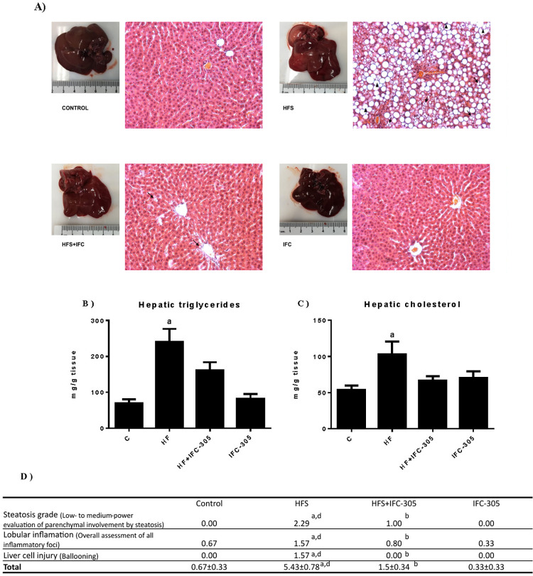
Fig 2. IFC-305 prevents alterations in the hepatic parenchyma and the development of steatosis.
(A) Representative liver sections from each experimental group were stained with H&E and observed at 20X magnification. Macrovesicular and microvesicular steatosis are indicated by ↑ and ▲, respectively. (B) Hepatic triglyceride and (C) cholesterol content. (D) H&E-stained liver sections observed at 20X magnification were analyzed according to semiquantitative Kleiner’s criteria. Statistically significant differences were determined by one-way analysis of variance (ANOVA) with multiple comparisons. “a” indicates a significant difference compared to the control group; “b” indicates a significant difference compared to the HFS group; “d” indicates a significant difference compared to the IFC group. A difference was considered significant when p≤0.05.

