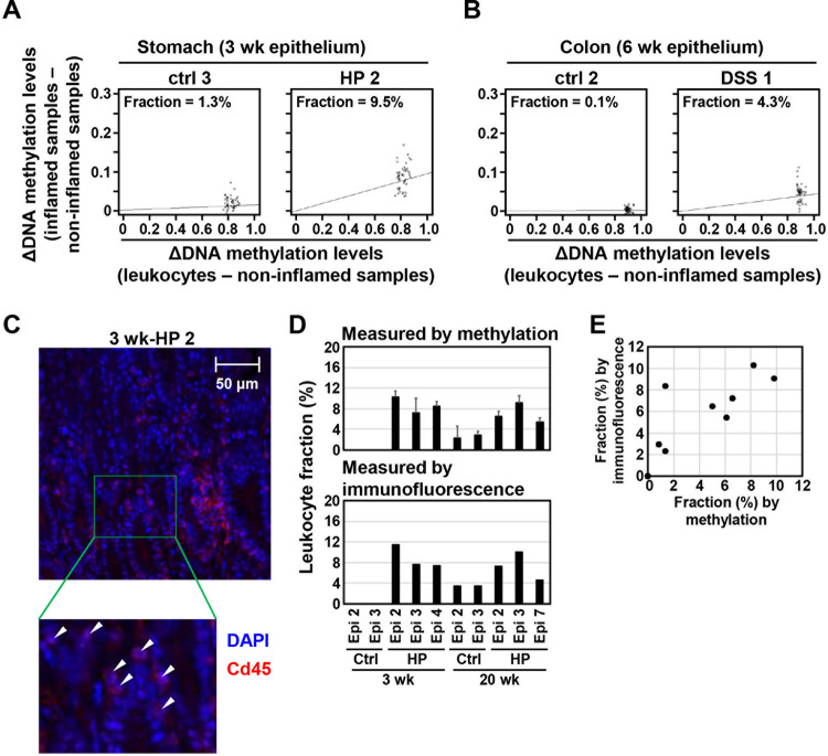
Fig 4. Estimation of leukocyte fraction in inflamed tissues by an algorithm using leukocyte-specific DNA methylation.
(A) and (B) Estimation of leukocyte fraction in H. pylori-infected stomach (A) and DSS-treated colon (B). The fraction of infiltrating leukocytes was 7.8% in H. pylori-infected stomach (HP 2) and 4.3% in DSS-treated colon (DSS 1), but 1.3% and 0.1% in their corresponding control samples (ctrl 3 and ctrl 2). (C) Histological estimation of a leukocyte fraction. The fraction of infiltrating leukocytes was histologically estimated by immunofluorescence of a leukocyte marker, Cd45, and DAPI staining. The number of DAPI-stained spots within Cd45-stained regions was counted as that of nuclei of leukocytes. White arrowheads show leukocytes. (D) and (E) The fraction of infiltrating leukocytes in H. pylori-infected stomach estimated using the algorithm with DNA methylation and that obtained histologically. The fraction in infected stomach was 7.6±3.6% and 7.8±1.6% using methylation markers and immunofluorescence, respectively (D). The fraction estimated using two methods was well correlated [R = 0.811 (p = 0.004)] (E).

