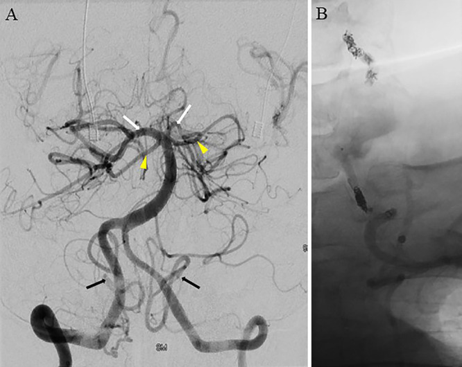
FIG. 3.

A: Right vertebral artery angiogram demonstrating robust filling of bilateral posterior cerebral arteries (white arrows), anterior inferior cerebellar arteries (arrowheads), and posterior inferior cerebellar arteries (black arrows). B: Proximal and distal coils occluding the left vertebral artery and trapping the aneurysm.
