Figure S1.
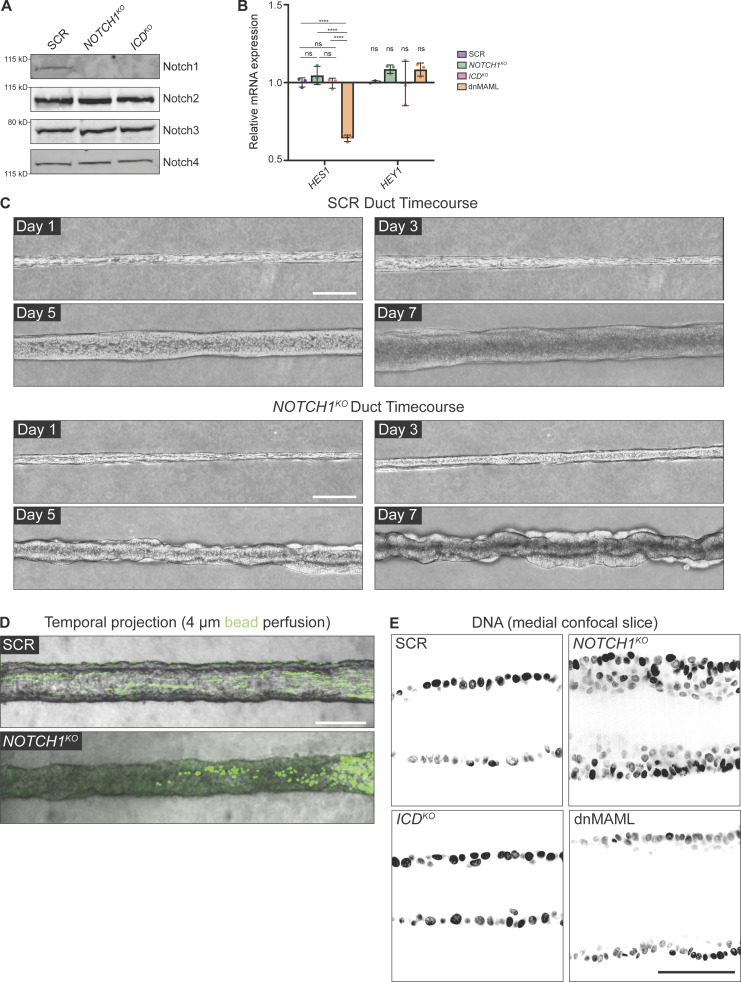
Notch1 cortical signaling influences mammary duct morphogenesis. (A) Western blot of lysates from scramble control (SCR), NOTCH1KO, and ICDKO cells immunoblotted for Notch1, Notch2, Notch3, and Notch4. (B) mRNA transcript expression of Notch1 target genes HES1 and HEY1 measured by qPCR in SCR, NOTCH1KO, ICDKO, and dnMAML-expressing cells. Average qPCR reads from n = 3 independent experiments. (C) Top: Phase contrast micrographs of SCR ducts over a 7-d timecourse shown at days 1, 3, 5, and 7 after seeding. Bottom: Phase-contrast images of NOTCH1KO ducts over a 7-d timecourse shown on days 1, 3, 5, and 7 after seeding. Scale bars, 150 µm. (D) Temporal projection micrographs of a timelapse of SCR and NOTCH1KO ducts perfused with 4 µm polystyrene beads (green). Scale bar, 200 µm. (E) Medial confocal slice micrographs from SCR, NOTCH1KO, ICDKO, and dnMAML ducts labeled with Hoechst (black). Scale bar, 50 µm. For plot B, mean ± SEM; one-way ANOVA with Tukey’s post-hoc test, ****P < 0.0001, ns denotes non-significant. Source data are available for this figure: SourceData FS1.

