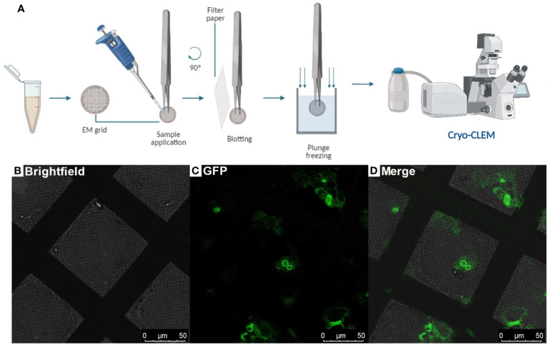
Figure 2.
Protoplasts vitrification by plunge-freezing and Correlative Light and Electron Microscopy (Steps 19 - 29 and 40 - 47) (A), Diagram showing the steps to follow after protoplast isolation: Plunge freezing procedure and observing the EM grids containing the root protoplasts under a cryo-CLEM, (B-D), Cryo-Fluorescence Microscopy of a grid containing root-protoplasts from Arabidopsis thaliana’s RAE1-GFP transgenic line. Brightfield: field of bright light; GFP: Green fluorescence signal (green); Merge shows the overlay of the brightfield and GFP signal.

