Figure 3.

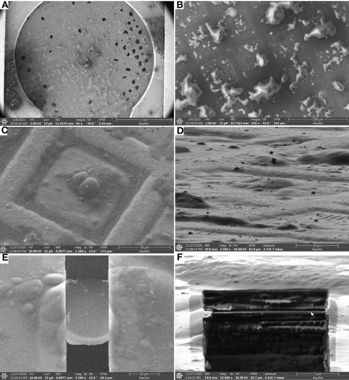
Lamella milling (Steps 48-56) (A), Scanning Electron Microscope (SEM) view of an EM grid containing vitrified root protoplasts from Arabidopsis thaliana RAE1-GFP transgenic line. (B, C), SEM images (top view) of root protoplasts before milling. (D), FIB view of root protoplasts. (E), SEM view of a cryo-FIB milled lamella, and (F), FIB view of a lamella. The lamella front in (F) is indicated by a white arrow. Scale bars: 1 mm (a), 200 µm (b), 50µm (c), 20 µm (d), 10 µm (E), and 5 µm (F).
