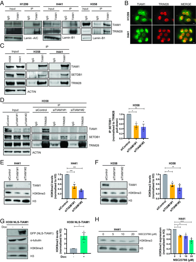
Fig. 4.
TIAM1 interacts with TRIM28 and SETDB1 to regulate H3K9 trimethylation. (A) Western blot analysis of endogenous TRIM28 co-immunoprecipitating with endogenous TIAM1 from the nuclear fraction of H1299, H441, and H358 cells. (B) Immunofluorescence images of H1299 and H441 cells stained for DNA with DAPI (blue), TIAM1 (green), and TRIM28 (red) with a merged overlay. (Scale bar, 20 μm.) (C) Western blot analysis of endogenous SETDB1 and TRIM28 co-immunoprecipitating with endogenous TIAM1. (D) Western blot analysis of endogenous SETDB1 co-immunoprecipitating with endogenous TRIM28, in cells transfected with TIAM1 or control siRNAs. The graph quantifies immunoprecipitated SETDB1 levels relative to immunoprecipitated TRIM28. (E and F) Western blot analysis of TIAM1 and H3K9me3 levels in H441 (E) and H358 (F) cells transfected with TIAM1 or control siRNAs. Graphs show H3K9me3 levels normalized to Histone H3. (G) Western blot analysis of NLS-TIAM1-GFP and H3K9me3 levels in uninduced control (−) H358 cells or cells with doxycycline-induced (+) expression of NLS-TIAM1. The graph shows H3K9me3 levels normalized to Histone H3. (H) Western blot analysis of H3K9me3 levels in H441 cells treated with an increasing dose of NSC23766. The graph shows H3K9me3 levels normalized to Histone H3. Data are presented as mean ± SEM of three independent experiments. *P < 0.05, **P < 0.01, and ***P < 0.001 (ANOVA).

