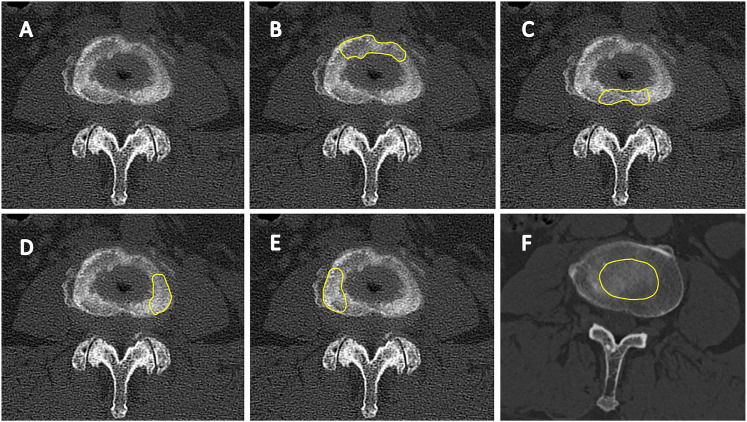
Figure 2.
(A) The epiphyseal ring on an axial computerized tomography image. (B) The ROI of the anterior epiphyseal ring. (C) The ROI of the posterior epiphyseal ring. (D) The ROI of the ipsilateral epiphyseal ring. (E) The ROI of the contralateral epiphyseal ring. (F) The ROI of the central endplate. ROI, region of interest.

