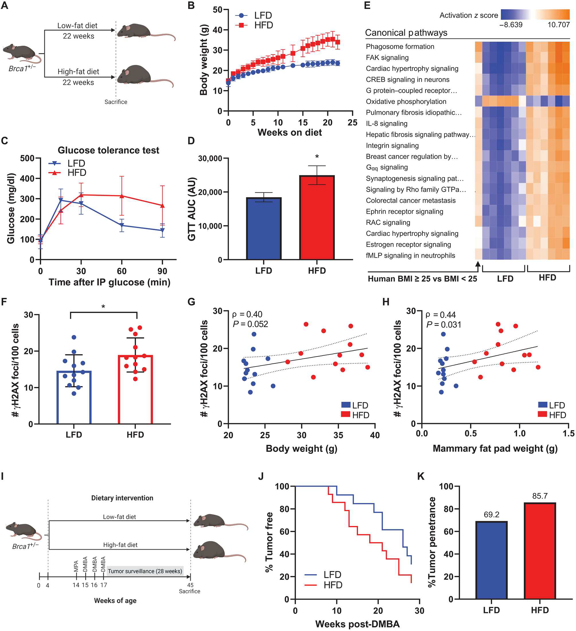
Fig. 6. HFD feeding leads to elevated mammary gland DNA damage in association with increased mammary tumor penetrance and decreased tumor latency in Brca1+/− mice.

(A) Experimental schematic of diet-induced obesity in C57BL6/J female Brca1+/− mice (n = 12 per group). (B) Average body weight of mice fed LFD or HFD over 22 weeks. (C) Glucose tolerance test conducted 1 week before euthanasia and (D) area under curve (AUC) calculation for each group (means ± SEM). AU, arbitrary units. (E) RNA-seq was conducted on whole mammary fat pad tissue from HFD and LFD mice (n = 6 per group). Activation of top 20 canonical pathways regulated in mammary fat pads from HFD mice compared with LFD mice is shown adjacent to corresponding pathway regulation in breast tissue from BRCA mutation carriers with BMI ≥ 25 versus carriers with BMI < 25 (n = 64 to 67 per group). (F) DNA damage assessed by IF (number of γH2AX foci/100 cells) in mammary glands at the time of euthanasia. (G) Correlation between mammary gland DNA damage and mouse body weight and (H) mammary fat pad weight among all mice. Spearman’s rank correlation coefficient (ρ) and associated P values are shown along with 95% confidence intervals. (I) Experimental schematic of medroxyprogesterone acetate/7,12-dimethylbenz[a]anthracene (MPA/DMBA)–induced tumorigenesis model in female Brca1+/− mice randomized to LFD or HFD groups (n = 13 or 14 per group). (J) Mammary tumor development in LFD and HFD mice shown as percentage of mice tumor-free over the 28-week surveillance period. (K) Overall mammary tumor penetrance at the end of the surveillance period shown as percentage of mice in each group that developed a mammary tumor. Student’s t test was used to determine significance unless otherwise stated. Data are presented as means ± SD unless otherwise stated. *P < 0.05.
