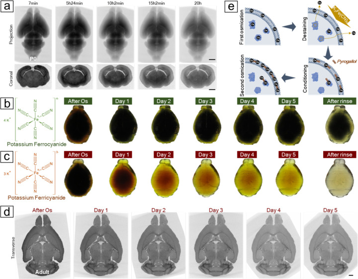
Fig. 2. Destaining the osmicated brain to remove excessive cytosolic osmium.
(A) Time-lapse X-ray microCT revealed a progressive decrease in X-ray attenuation from an osmicated P0 mouse brain after the application of potassium ferrocyanide. (B) Destaining a protein-infused mouse brain proxy with ferrocyanide showed that after five days treatment, only the superficial layer was efficiently destained. (C) The brain proxy treated with ferricyanide became clear after five days of treatment. As ferrocyanide turned dark green and ferricyanide remained yellow during the reaction, a thorough rinse was applied after five days to reveal the actual color of the brain proxies. (D) A virtual transverse section from the X-ray microCT reconstruction of a mouse brain treated with ferricyanide displayed a decrease in X-ray attenuation, especially in gray matter areas, which enhanced the contrast of the white matter tracts over the course of the reaction. (E) A reaction mechanism was proposed based on the X-ray observations and the brain proxy tests. Initial osmication with OsO4 stains both cell membranes and cytoplasm. Cytoplasmic osmium bound to proteins can be stripped and washed off using ferricyanide, while the remaining osmium anchors multidentate ligands, such as pyrogallol, which provide more binding sites for the second round of osmium binding. Scale bars: 1 mm.

