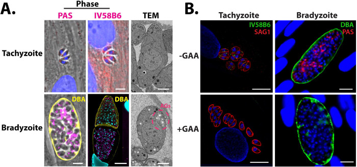
Figure 1.
Glucan dynamics in T. gondii ME49 tachyzoites and in vitro bradyzoites.
A, Microscopy-based glucan evaluation of T. gondii tachyzoites and bradyzoites using PAS (left), IV58B6 (middle; α-glycogen IgM mAb), and TEM (right). B, GAA digest of AGs in tachyzoites and bradyzoites confirms specificity of IV58B6 antibody and PAS staining. All scale bars = 5 μm.

