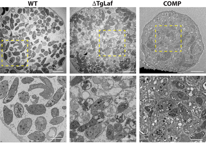
Figure 9.
Transmission electron microscopy of purified tissue cyst from infected mouse brains confirms the presence of aberrant AG accumulation within ΔTgLaf cysts. The accumulation of amylopectin granules within the cytoplasm of WT bradyzoites with parasites exhibiting different levels. Additionally, evidence of active endodyogeny is present. ΔTgLaf tissue cysts contain a mix of bradyzoites with expected cytoplasmic and organellar contrast as well as others with grossly exaggerated AG levels that and the apparent loss of both the cytoplasmic and organellar contents. The COMP line exhibits AG accumulation levels similar to that observed in WT parasites with the tissue cyst itself appearing to be very tightly packed, with high levels of granular material between individual bradyzoites. Upper panels: scale bar = 5 μm; lower panels (zoom of boxed region from upper panel): scale bar = 2 μm.

