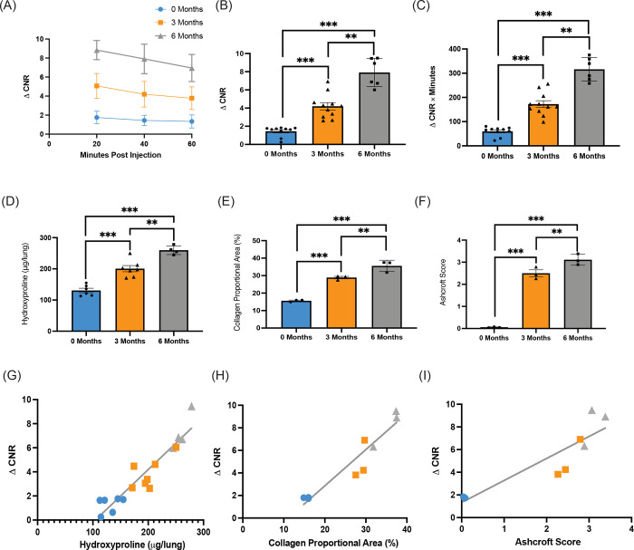
Figure 2:
EP-3533 detects progressive RILI in murine model. ΔCNR versus time curves show progressively increased EP-3533 enhanced MR signal at 3 months and 6 months post irradiation (A). ΔCNR at 40 minutes post-injection is progressively elevated from 0 months, 3 months, and 6 months post irradiation (B). Area under the curve quantification also demonstrated similar elevations in EP-3533 signal (C). Validated techniques used to quantify RILI include hydroxyproline (D), collagen proportional area (E), and Ashcroft score (F). Each technique demonstrates increasing severity of RILI over time post irradiation with ANOVA and Bonferroni post hoc t-test. The association between EP-3533 and hydroxyproline (G), collagen proportional area (H), and Ashcroft score (I) were assessed via Pearson correlation and EP-3533 was significantly correlated with RILI severity in each. (* p<0.05, ** p<0.001, *** p<0.0001).

